High Efficient Isolation and Characterization of Endothelial Cells from Newborn Cord Blood Veins
Manizheh Azhdari, Hossein Baharvand, Mohamadreza Baghaban- Eslaminejad and Nasser Aghdami
1 Department of Stem Cells and Developmental Biology, Cell Science Research Center, Royan Institute for Stem Cell Biology and Technology, ACECR, Tehran, Iran
2 Department of Developmental Biology, University of Science and Culture, ACECR, Tehran, Iran
3 Department of Regenerative Medicine, Cell Science Research Center, Royan Institute for Stem Cell Biology and Technology, ACECR, Tehran, Iran
- *Corresponding Author:
- Baharvand H
Department of Stem Cells and Developmental Biology
Cell Science Research Center
Royan Institute for Stem Cell Biology and Technology
ACECR, Tehran, Iran.
Tel: +00982126300025
Email: Baharvand@RoyanInstitute.org - Aghdami N
Department of Stem Cells and Developmental Biology
Cell Science Research Center
Royan Institute for Stem Cell Biology and Technology
ACECR, Tehran, Iran.
Tel: 982122339936
Email: Nasser.Aghdami@RoyanInstitute.org
Received date: July 11, 2016; Accepted date: July 30, 2016; Published date: August 05, 2016
Citation: Azhdari M, Baharvand H, Ghasemzadeh-Hasankolaei M, et al. High Efficient Isolation and Characterization of Endothelial Cells from Newborn Cord Blood Veins. J Clin Dev Biol. 2016, 1:3.
Abstract
This study introduces a fast and efficient protocol for isolation of human vascular endothelial cells from veins of newborn cord blood for using in cell and drug therapy. Endothelial cells act as barrier between blood and lower layers. These cells, because of their tight connections allow selective molecules to pass through them and loss of them by several factors causes various diseases in humans. Because many studies are not possible in a living organism, hence isolation of consider cells in vitro are also key factors for in vitro studding.
Here in, we have introduced an efficient protocol for isolation and culture of endothelial cells from newborn cord blood veins to achieve high purity endothelial cells (above 98%) for in vitro studding, drug and therapy using. This protocol comprises enzymatic and mechanic cord blood veins dissociation as well as a purification step process using anti-CD31 antibodies conjugated to MACS, which produces a pure endothelial cell population of newborn cord blood veins.
Keywords
Endothelial cells; Isolation; Cord blood vein
Introduction
The endothelium is a layer that covers the internal surface of blood vessels. It consists of 3 layers and the innermost layers a row of endothelial cells. Vascular endothelial cells entire circulatory system from heart to capillaries and play a major role in tissue homeostasis. These cells responses involve barrier between lumen and rounding tissues, blood clotting, angiogenesis, vasculogenesies and their important role in body is filtration function [1,2]. Loss and dysfunction of endothelial cells causes hypertension, thrombosis and cardiovascular failure and also a broad spectrum of diseases such as atherosclerosis and lesion formation. Most of our knowledge from these cells has led to the further our understanding of these cells dysfunction or integrity in vivo [3].
Asahara et al., was the first who discovered the existence of Endothelial Progenitor Cells (EPCs) in human peripheral blood and he believed that EPCs originate from the bone marrow [4]. But the number of EPCs is very low and is not be able to repair damaged vessels [5]. Assessment of endothelial function may be a useful tool for coronary artery disease. In addition, involving the mechanisms and molecular pathways in endothelial cells can be further elucidated through studying of these cells in vitro. ECs play a major role in tissue homeostasis and in diverse pathologies. A pure source of isolated these cells can allow to study endothelial cells role in the formation of vessels and pathogenesis of vascular diseases. There are some methods that isolate ECs only from the eyeball [6], lung [7,8] or murine heart [7,8]. These vessels are not as easily obtained as umbilical cords. A number of investigators have been attempted for culture of endothelial cells over the last few years [9,10] but they have 2 problems: a) Endothelial cells maintain inability in pure culture for long time and b) Identification inability of the cultured cells.
Here, we present a protocol for isolation and characterization of Endothelial Cells (ECs) from Human Umbilical Vein (HUVEC) using enzymatic (collagenase type IV) and after their confluence mechanical purification (CD31-antibody FACS). Because these cells are easy to obtain, culture and can be reproduce high number during limited time in comparison with other protocols.
Materials and Methods
Antibodies
Monoclonal antibodies for human CD31 (1:200, 555444), vWF (1:200, 555849), VE-cad (1:200, 555661) for immunostaining and were purchased from Becton Dickenson (BD).
Secondary murine polyclonal antibodies IgG1-FITC (1:200, 04611) and IgG1-PE (1:200, 340270) were obtained from BD.
Media, growth factors, enzymes and supplements
Medium M-199 (Gibco-Invitrogen, 31100-027)
Fetal Bovine Serum (FBS) 10% (Gibco-Invitrogen, 10270106)
L-glutamine 2 mM (Gibco-Invitrogen, 25030-024)
Nonessential Amino Acid solution 1% (NEAA) (Gibco-Invitrogen, 11140-035)
Penicillin-streptomycin 1% (pen/strep) (Gibco-Invitrogen, 15070- 063)
Phosphate Buffered Saline (PBS) (Gibco-Invitrogen, 21600-069)
Collagenase IV (0.1%, 1 mg/ml)
Trypsin–EDTA 0.05% (Gibco-Invitrogen, 15400-054)
Cell and molecular biology reagents and kits
Taq DNA polymerase (Takara, RP01AM)
Ethidium bromide (Sigma-Aldrich, E7637)!
Caution! Ethidium bromide is carcinogenic. Dispose of all contaminated gels, buffers and tips. Also, wear gloves.
Agarose (Sigma-A2278)
Trizol (Invitrogen, 10296-028)!
Caution! Trizol is carcinogenic. Use gloves and work under a hood.
Primers for RT-PCR (Table 1)
| GENE TRANSCRIPT | PRIMER SEQUENCES | PRODUCT(BP) |
|---|---|---|
| GAPDH | F:5-CTCATTTCCTGGTATGACAACGA-3 R:5-CTTCCTCTTGTGCTCTTGCT-3 |
122 |
| CD31 | F-5-AGCAGTACCACTTCTGAACTCC-3 R-5-AGGAATTGCTGTGTTCTGTGG-3 |
428 |
| VE-cadherin | F-5-CTCCAACTCCATACTCCACTC-3 R-5-AGTCTCAAAGCAAGGTCTCAG-3 |
319 |
| vWF | F-5-CATCTAGCTAAGAGGAGGAC-3 R-5-TTGTGTTCATCAAAGGGTGG-3 |
150 |
Table 1: PCR primer sequences for marker genes.
Paraformaldehyde (PFA) 16% (wt/vol) solution (Sigma- Aldrich-P6148)
Formalin solution, neutral buffered 10% (Sigma-F8775)!
Caution! PFA and formalin solution is toxic. Use gloves work under a hood!
Triton X-100 (1.00014).
4′,6-Diamidino-2-phenylindole (DAPI, 1:1000 Sigma-Aldrich, D-8417).
DNase I (RR066A).
Equipment
FACS tubes (BD, 352235)
15 ml Falcon tubes (BD, 357551)
500 ml Medium filtration systems (BD, 212351)
50 ml Medium filtration systems (BD, 430758)
Plastic disposable pipettes, 1, 5, 10, 25 and 50 ml
Tissue culture incubator, 5% CO2/95% air, 37°C
PCR thermocycler
Vortex
Flow cytometry cell sorter: FACStar with CellQuest software or FACSAria with FACSDiva software (FACS Aria, USA)
Collagenase type-IV solution: Dissolve (0.1%) 1 mg collagenase in 1 ml DMEM. Filter the solution using a 50 ml filtration system and store at 4°C. The Solution can be used up to 2 weeks.
FACS buffer Add 2% (vol/vol) FBS and 1% (vol/vol) pen strep to PBS and filter (0.22 μm filter). Store at 4°C up to 2 weeks
DNase solution (100 mg) in 10 moldable distilled sterile water. Aliquot and store at -20°C up to 6 months.
4% PFA Dilute 10 ml 16% (wt/vol) PFA in 40 ml PBS. Aliquot and store at -20°C.
0.2% Triton X-100 solution Add 200 μl Triton X-100 to 100 ml PBS and mix thoroughly. Store at room temperature (22-25°C).
DAPI solution Dilute DAPI 1:1,000 in PBS as follows: Pipette 1 μl DAPI into 1 μl PBS and mix well. Add 5 μl PBS. 1 μl after another, and mix thoroughly. Then gradually add 994 μl PBS. Prepare the dilution fresh, on ice and in the dark.
Reagent setup
EC medium: Add 250 μl VEGF solutions to 50 ml EGM-2 medium (final concentration of VEGF is 50 ng/ml). Store at 4°C up to 2 weeks.
Collagenase type-IV solution: Dissolve 100 mg collagenase in 50 ml DMEM. Filter the solution using a 50 ml filtration system and store at 4°C. The solution can be used up to 2 weeks.
FACS buffer: Add 5% (vol/vol) FBS and 1% (vol/vol) pen/strep to PBS and filter (0.22 μm filter). Store DNase solution: Reconstitute lyophilized DNase (100 mg) in 10 ml double distilled, sterile water. Aliquot and store at -20°C up to 6months.
PFA: Dilute 10 ml 16% (wt/vol) PFA in 40 ml PBS. Aliquot and store at -20°C.
0.2% Triton X-100 solution Add 200 μl Triton X-100 to 100 ml PBS and mix thoroughly. Store at room temperature (22-25°C).
DAPI solution: Dilute DAPI 1:1,000 in PBS as follows: pipette 1 μl DAPI into 1 μl PBS and mix well. Add 5 μl PBS. 1 μl after another, and mix thoroughly. Then gradually add 994 μl PBS.
CAUTION! Prepare the dilution fresh, on ice and in the dark.
Equipment setup
FACS setup FACSAria equipped with FACSDiva software is used for cell sorting and analysis.
Set FACS parameters: Set 585-nm for PI detection, and 530-nm band pass filter for FITC detection. Use a wide nozzle of 100 μm. Use control samples to determine appropriate settings.
For isotype control-use anti-IgG1κ-FITC antibody.
For positive control-use ECs, such as HUVEC, that express CD31.
For Negative control, to assess the background autofluoresence of cells-no antibody is placed over samples.
For Cell viability control, that can help in identifying and excluding dead cells from sorted cells-use PI (0.5 μg/ ml) staining to detect dead cells.
Cell Characterization
Immunostaining
Cells were fixed in 4% paraformaldehyde, blocked by 1% rat serum (Royan Institute), and then incubated overnight with primary antibodies at 4°C. Anti-mouse IgG antibodies conjugated with PE or FITC were used as secondary antibodies. Nuclei were stained with DAPI (1:1000 Sigma-Aldrich, D-8417). After each incubation, cells were washed twice with PBS. Stained cells were photographed with fluorescence microscopy (BX51; Olympus; Japan).
Gene expression analysis
Total RNA from 1 × 10⁶ ECs were extracted with using Trizol according to the manufacturer’s protocol.
Total RNA can be stored at -80°C until use.
Reverse transcription (RT) reaction was carry out with 1 μg extracted RNA using the RT enzyme kit and according to manufacturer’s protocol. cDNA can be stored at -80°C until use.
PCRs with DNA polymerase Performed with using 1 μl of RT product per reaction.
Glyceraldehyde-3-phosphate dehydrogenase (GAPDH) was used as an internal standard.
Flow cytometry and cell sorting analyses
Dissociation of cells was performed with trypsin/EDTA (Gibco- Invitrogen, 15400-054). Following neutralization with M-199 supplemented with 10% FBS, cells were washed in PBS that contained 2% FBS (FACS buffer). Subsequently, cells were incubated for 1 h at 4°C with mouse IgG1 monoclonal anti-human CD31 antibody and then washed with FACS buffer. Secondary polyclonal rat anti-mouse IgG1-PE or FITC was added and cells incubated for 1h at 4°C (Table 2). After washing with FACS buffer, CD31-positive cells weresorted by FACS Calibur (USA). Cells were fixed with 4% paraformaldehyde) Sigma-Aldrich, P6148).
| Antibody | Company | Cat.N |
|---|---|---|
Mouse monoclonal anti-human CD31 |
BD | 555444 |
Mouse monoclonal anti-human vWF |
BD | 555849 |
Mouse monoclonal anti-human VE-cad |
BD | 555661 |
Rat anti-mouse IgG1-FITC |
BD | 04611 |
Rat anti-mouse IgG1-PE |
BD | 340270 |
Table 2: Antibody sources.
Uptake of acetylated low-density lipoprotein (DiI-ac-LDL)
Acetylated Low-Density Lipoprotein (DiI-ac-LDL; Biomedical Technologies Inc., MA) was diluted to 10 μg/ml in complete growth medium and added to the cells and the mixture incubated for 4 h at 37°C. Then, the medium was removed and washed with probe-free media. Cells fixed with 4% paraformaldehyde. The nuclei were stained with 4,6-diamidino-2-phenylindole (DPAI, 1:1000 Sigma-Aldrich, D-8417) and finally visualized with fluorescence microscopy.
Tube formation by ECs in vitro
Endothelial cellswere cultured to form tubes in vitro as described previously. Briefly, Matrigel matrix was diluted to 0.5-0.7 mg/ ml in M-199 medium. ECs trypsinized and replated at 3-5 × 10⁴ cells in 300 μl of 0.5 - -0.7 mg/ml Matrigel onto each well of the 12-well plates. The Matrigel-cell suspension polymerized for 2-4 h at 37°C. Again, 300 μl of M-199 medium was added by semi-depletion to the dish and cells were incubated at 37°C and 5% CO2. The structures photographed under phase contrast microscope (Olympus, IX71).
Procedure
This procedure describes the isolation of endothelial cells from human umbilical cord blood using CD31-antibody (Figure 1).
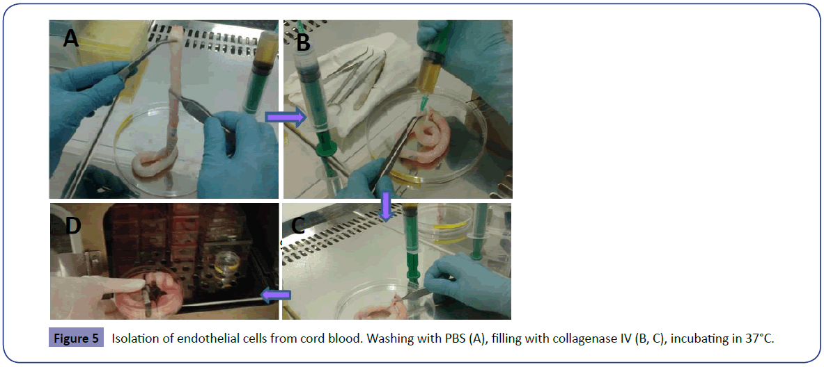
Figure 1: Schematic illustration of the isolation of endothelial cells from human new born umbilical cord blood. After washing with PBS, cord blood were filled with collagenase IV and incubated in 37°C. After cell isolation, culture and confluenceing, CD31 positive cells were isolated with FACS anad re cultured in M199 medium.
Isolation of endothelial cells from newborn cord blood
Wash umbilical cords from newborn infants with phosphate buffered saline to remove internal blood. Then, fill veins with collagenase IV and incubated at 37°C for 10 min. After massaging the cord blood, collecting the cell suspensions and centrifuging at 4°C and 1500 rpm, culture cell pellets in M-199 medium supplemented with 10% FBS, 1% NEAA, and 1% penicillin/ streptomycin (TIMING: 30 min).
Critical step! Be careful during massaging, it done very slowly, otherwise the fibroblasts also separate from tissue and ECs purity will be low.
Isolation of CD31+ cells from cultured cells
After reaching confluence, the Cells were dissociated using trypsin/EDTA.
Then, cells were washed with PBS and stained with monoclonal antibodies for CD31. After washing with FACS buffer, CD31- positive cells were sorted by FACS Calibur (USA) (TIMING: 120 min).
Statistical Analysis
We used repeated measures ANOVA followed by the Tukey post hoc test. Data were expressed as mean ± SD. Repeated number were n ≥ 3.
Results
This protocol presents a simple method for isolation of ECs from human umbilical vein, with a typical yield of above 98% CD31- positive cells after FACS sorting during 5-7 days.
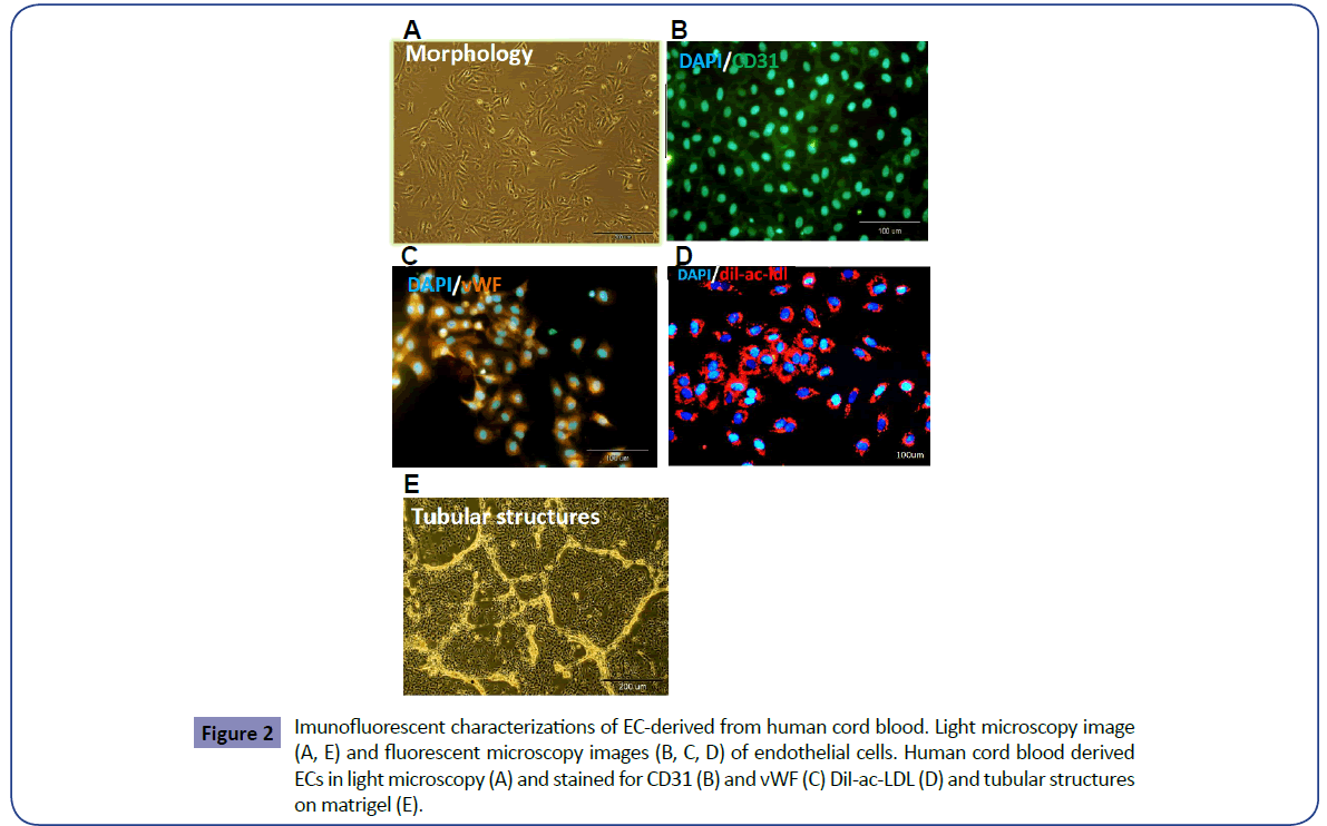
The isolated and cultured CD31-positive cells show cobblestonelike cell phenotypic endothelial characteristics (Figure 2A). High purity of nearly 98% CD31-expressing cells resulted when assessed with Flow cytometry, DiI-labeling and CD31 immunostaining after FACS sorting (Figures 2B and 2C).
Figure 2: Imunofluorescent characterizations of EC-derived from human cord blood. Light microscopy image (A, E) and fluorescent microscopy images (B, C, D) of endothelial cells. Human cord blood derived ECs in light microscopy (A) and stained for CD31 (B) and vWF (C) DiI-ac-LDL (D) and tubular structures on matrigel (E).
CD31+cells isolated in this manner showed the endothelial bio functional characteristics by efficiently incorporating DiI-ac-LDL as detected by fluorescence microscopy after incubation with this substance (Figure 2D).
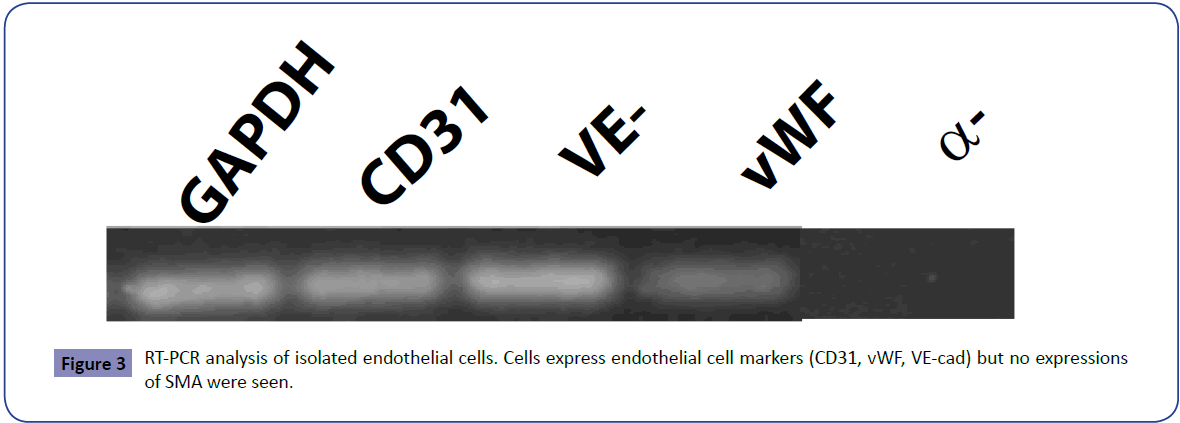
The endothelial cells isolated and expanded in vitro express similar levels of typical endothelial, such as CD31, VE-cad and vWF genes but these cells didn’t express α-SMA (Figure 3).
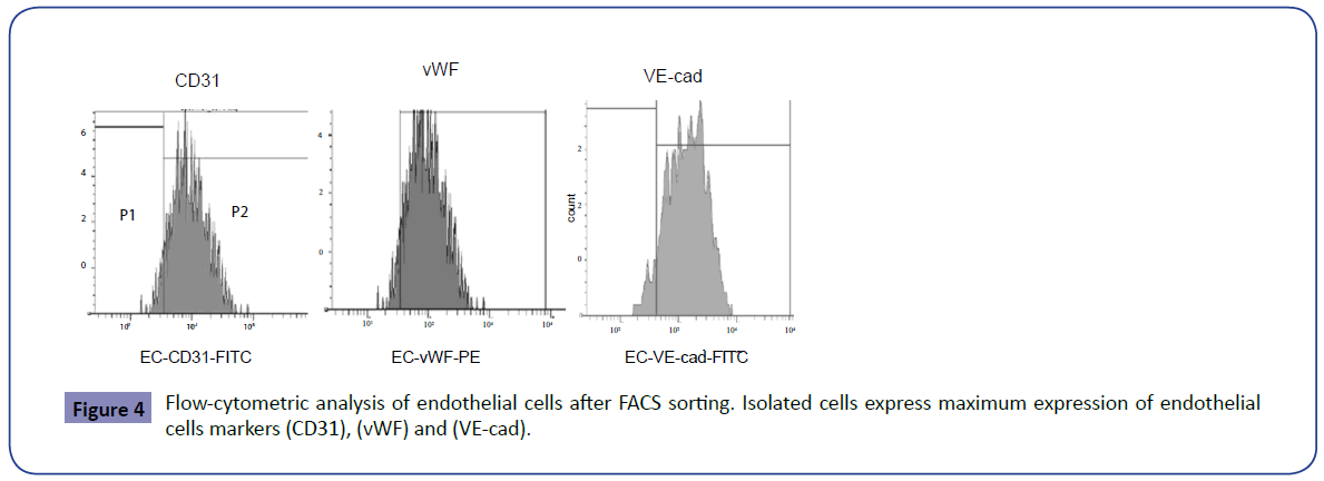
Flow cytometry analyses revealed the isolated and sorted-ECs expressed similar levels of CD31 ( ̴97%), vWF ( ̴95%) and VE-cad ( ̴96%) (Figure 4).
Effective incorporation of DiI-ac-LDL and formation of blood vessel-like structures in vitro by Matrigel were used to determine ECs functionality (Figure 2D and 2E).
Discussion
Endothelium dysfunction, loss of endothelial cells and also these cells abnormal functionality are the main aspects of vascular diseases and are regarded as key events in development of some related diseases such as atherosclerosis, hypertension, thrombosis, diabetes mellitus and hypercholesteremic [2,11,12]. The main functions of endothelial cells include participating in: coagulation, immune function, platelet adhesion and also volume controlling of extravascular and intravascular spaces [2,13,14].
A further consequence of damage to the endothelium is related of some factors, which promote platelet aggregation and adhesion to the sub endothelium and inflammatory responses [15,16].
For understanding how the vascular dysfunction development and the initiators of endothelial injury, it is necessary to consider the normal biology of the endothelium, endothelial cells functions, biological molecules under its control, and also assessment specific processes that are deregulated in related diseases.
in vitro isolation and studying of Endothelial Cells (ECs) is an invaluable tool by which the function of the vascular endothelium in normal physiology and patho-physiology can be prospect.
In the present study we describe a simple protocol for the isolation of endothelial cells from human newborn cord blood vein. The protocol involves enzyme digestion and positive selection with anti-CD31 Abs and FACS sorting. Our results show that the isolation protocol with enzymes and FACS sorting was relatively efficient. After enzyme digestion, cell culturing and FACS sorting with CD31 ABs, the purity and functional properties of the isolated cell lines were extensively characterized. The isolated cells showed maximum expression of CD31, vWF, and VEcad in flow cytometry analysis (Figures 4 and 5). The presence of smooth muscle cells was also excluded after FACS sorting; these cells in RT-PCR analysis were negative for SMA (Figure 3). Finally, isolated endothelial cells functionality demonstrated their ability to form capillary-like structure on Matrigel and also capable of uptakeing DiI-ac-LDL (Figures 2D and 2E). In this procedure, high purity, good functionality, efficient passaging and also cells viability are great interest. The morphology and phenotype of these cultured cells remain stable over 20 passages in culture.
This protocol outlined here is to isolate and culture ECs in simple and effective way for scientific and medical studding. We conclude that the protocol described herein allows purification and culture of ECs from human newborn cord blood with very high efficiency and longtime maintains.
Acknowledgments
This study was funded by a grant provided by the Royan Institute.
References
- Verma S, Anderson TJ(2002) Fundamentals of endothelial function for the clinical cardiologist. Circulation 105: 546-549.
- Deanfield J, Donald A, Ferri C, Giannattasio C, Halcox J,et al. (2005) Endothelial function and dysfunction. Part I: Methodological issues for assessment in the different vascular beds: a statement by the Working Group on Endothelin and Endothelial Factors of the European Society of Hypertension. J Hypertens23: 7-17.
- Zeiher AM, Drexler H, Wollschläger H, Just H (1999) Endothelial dysfunction of the coronary microvasculature is associated with impaired coronary blood flow regulation in patients with early atherosclerosis. Circulation 84: 1984-1992.
- Asahara T, Murohara T, Sullivan A, Silver M, van der (1997)Isolation of potative progenitor endothelial cells for angiogenesis. Science 275: 964-967.
- Asahara T (2007) Cell therapy and gene therapy using endothelial progenitor cells for vascular regeneration. HandbExpPharmacol180: 181-194.
- Su X, Sorenson CM, Sheibani N (2003) Isolation and characterization of murine retinal endothelial cells. Mol Vis9: 171-178.
- Marelli-Berg FM, Peek E, Lidington EA, Stauss HJ, Lechler RI (2000) Isolation of endothelial cells from murine tiss. J Immunological Methods 244: 205-215.
- Lim YC, Cardena GG, Allport JR, Zervoglos M, Connolly AJ, et al. Heterogeneity of Endothelial Cells from Different Organ Sites in T-Cell Subset Recruitment. AmJ Pathol 162: 1591-1601.
- Maruyama Y (1963) The human endothelial cell tissue culture. Z ZellforschMikrosk A4nat 60: 69-79.
- Pollak OJ, Kasai T (1964) Appearance and be-havior of aortic cells in vitro. Amn J AMcdSci248: 71-78.
- Münzel T, Sinning C, Post F, Warnholtz A, Schulz E(2008) Pathophysiology, diagnosis and prognostic implications of endothelial dysfunction. Ann Med 40:180-196.
- Flammer AJ, Anderson T, Celermajer DS, Creager MA, Deanfield J (2012) The assessment of endothelial function: from research into clinical practice. Circulation 126:753-767.
- Atkinson G, BatterhamAM (2013) Response to “Adjusting for brachial artery diameter in the analysis of flow-mediated dilatation: Pitfalls of a landmark paper?”Atherosclerosis 228: 282-283.
- Gokce N(2011) Clinical assessment of endothelial function: ready for prime time? CircCardiovasc Imaging 4: 348-350.
- Roberts OL, Holmes K, Müller J, Cross DA, Cross MJ (2009) ERK5 and the regulation of endothelial cell function. BiochemSoc Trans 37: 1254-1259.
- Suessenbacher A, Potocnik M, Dörler J, Fluckinger G, Wanitschek M, et al.(2011) Comparison of peripheral endothelial function in shift versusnonshift workers. Am J Cardiol107:945-948.
Open Access Journals
- Aquaculture & Veterinary Science
- Chemistry & Chemical Sciences
- Clinical Sciences
- Engineering
- General Science
- Genetics & Molecular Biology
- Health Care & Nursing
- Immunology & Microbiology
- Materials Science
- Mathematics & Physics
- Medical Sciences
- Neurology & Psychiatry
- Oncology & Cancer Science
- Pharmaceutical Sciences

